Active
Clinical Validation Toolkit
Calibration, decision-curve and external-validation checks packaged so a model can be re-tested on a new cohort without rebuilding the evaluation stack.
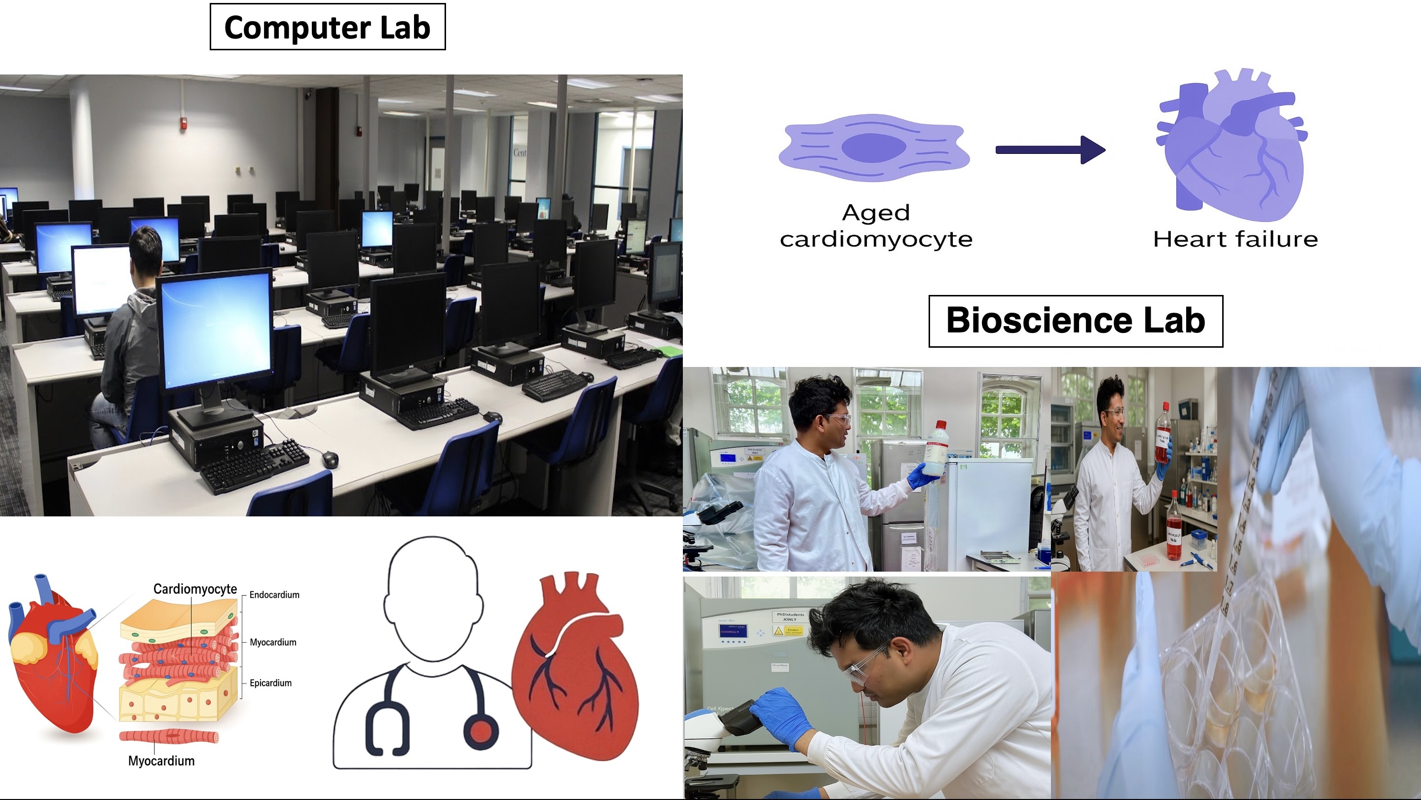
We run two complementary spaces — a Computer Lab and a Bioscience Lab — to build interpretable, clinically useful models for cardiovascular health. Our flagship project is CardioFlowFormer, a spatiotemporal framework for motion phenotyping and early risk detection.
Our Labs
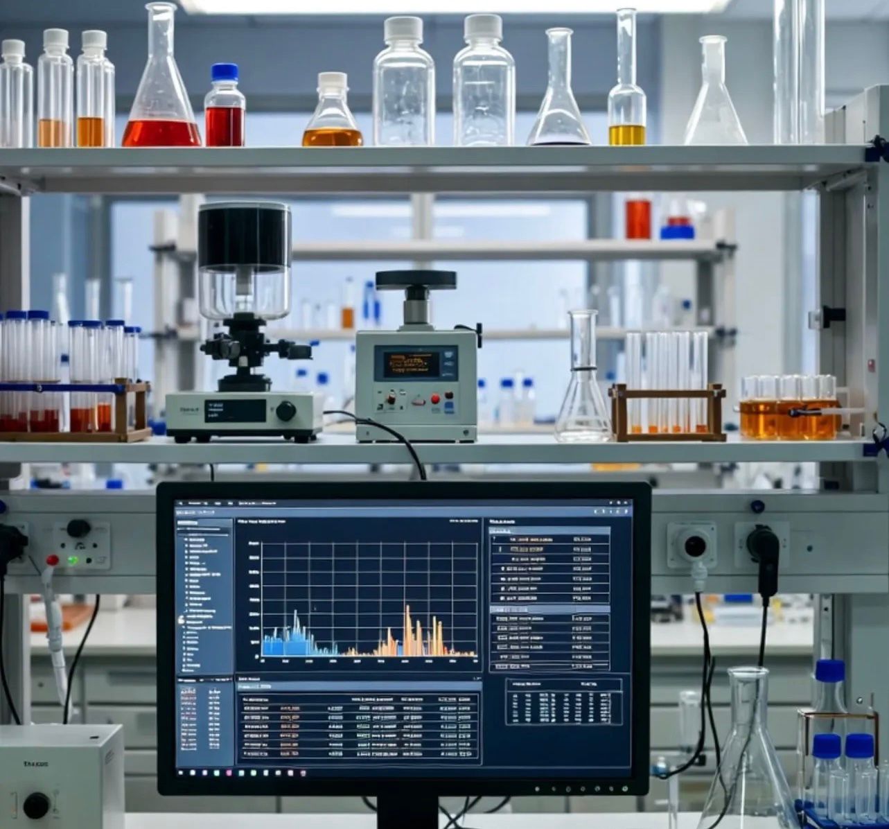
Cells are imaged next door to where the models are trained, so a labelling problem at the microscope reaches the pipeline the same week it appears.
Computer Lab
Bioscience Lab
Flagship model
Transformer-based spatiotemporal model that fuses motion fields, morphology, and temporal contraction metrics to detect ageing- and damage-related cardiomyocyte dysfunction early.
How it works
Four stages, each one auditable on its own. The motion field and the attention weights are kept, so a prediction can be traced back to the frames that produced it.
Step 01
iPSC-derived cardiomyocytes are filmed beating in culture, with annotations made at the microscope.
Step 02
Frame-to-frame displacement is estimated across the cell, turning contraction into a dense motion field.
Step 03
Flow patches become tokens; temporal attention weighs which regions and which beats carry the signal.
Step 04
Motion phenotypes feed calibrated time-to-event models, with attention maps retained for inspection.
Active Projects
Calibration, decision-curve and external-validation checks packaged so a model can be re-tested on a new cohort without rebuilding the evaluation stack.
Subgroup performance and bias auditing for cardiovascular risk models, reported across age, sex and ethnicity rather than as a single pooled AUC.
Optical-flow feature extraction from cardiomyocyte video, turning contraction dynamics into quantitative phenotypes for downstream modelling.
People
Founder, Cardio AI Lab
Research: spatiotemporal modelling, survival analysis, fair & interpretable AI for CVD.
Email: m.sufian@uel.ac.uk
Bioscience Lab · Clinical partners
Bioscience Lab (UEL), clinical partners, and external collaborators across imaging and biostatistics.
Join / Collaborate
We welcome collaboration on datasets, clinical validation, and tooling. Email m.sufian@uel.ac.uk or m.sufian@bbk.ac.uk.